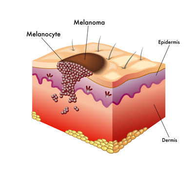
An abnormal looking mole should be biopsied by a physician and sent to a pathologist. The pathologist will look at the mole under the microscope and determine if it is melanoma or not. Once the diagnosis of melanoma has been made, your doctor and the pathologist will determine the stage of the disease.
To find out whether the melanoma has spread or not, a physician will feel the lymph nodes around the area of the melanoma. The doctor may order a test called a sentinel node biopsy in order to see if the tumor has spread to the lymph nodes closest to the melanoma. If the lymph node is involved with melanoma, the doctor may recommend an operation to remove all of the lymph nodes in the area. If this is the case, additional tests such as a CT scan or PET scan may be ordered to be sure that the tumor has not spread to other parts of the body.
Melanoma can be classified according to four stages, from I to IV, indicating the severity of the disease.
- Patients with stage I and II disease have melanoma in the skin only. Patients are classified as Ia, Ib, IIa, IIb or IIc depending on how deep the melanoma goes into the skin (Breslow’s thickness) and whether the melanoma has an ulcer or not in its surface.
- Patients with stage III disease have melanoma that has spread to the lymph nodes.
- Patients with Stage IV disease have melanoma that has spread to other parts of the body such as the liver, lung or brain.
Causes of Melanoma
Although few studies, mostly in Germany and Australia, have shown that most children with melanoma have some of the same risk features as adults, there are other conditions that increase the chances of melanoma in children. Risk factors for children include:
- The presence of melanoma at birth (congenital melanoma) as a result of passage of the tumor from the mother to the infant through the placenta
- The presence of a giant mole
- Diagnosis of a rare disorder called xeroderma pigmentosum
- Presence of a rare disease called neurocutaneous melanosis
- Werner’s syndrome
- History of the genetic form of retinoblastoma
- A weakened immune system after bone marrow or kidney transplant or as a result of an infection such as HIV